
Figure 1.
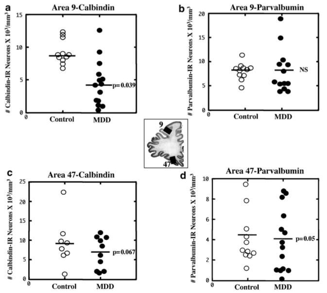
Comparison of the cell packing density of CB-IR (a and c) and PV-IR (b and d) neurons in Brodmann's areas 9 and 47 (see the middle picture for the localization of these areas) of control subjects and subjects with MDD. A significant 50% reduction in the density of CB-IR neurons in Brodmann's area 9 in MDD subjects (a) and a 27% trend toward a reduction in CB-IR (c) and PV-IR neurons (d) in Brodmann's area 47 are illustrated. Values for the individual subjects (circles) and mean values (horizontal lines) are given without the adjustment for the covariates.