
Figure 2.
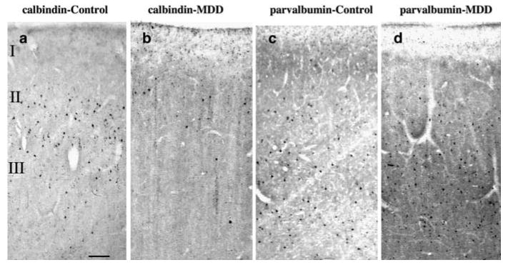
Photomicrographs of one pair of age-matched control and MDD subjects showing CB (a and b) and PV (c and d) immunoreactive neurons in the upper two-thirds of the cortical width in Brodmann's area 9 of the dlPFC. Note that CB-IR neurons are localized in upper cortical layers II + IIIa (a and b), whereas PV-IR neurons are more numerous and have a wider distribution across middle and lower cortical layers III–VI (c and d). Although adjacent sections from area 9 were used for each of the markers, there is a 50% reduction in CB-IR neurons in MDD as compared to control subjects, whereas no differences in PV-IR neurons were observed between the groups. Images were obtained using the × 4 objective, scale bar = 125 μm.