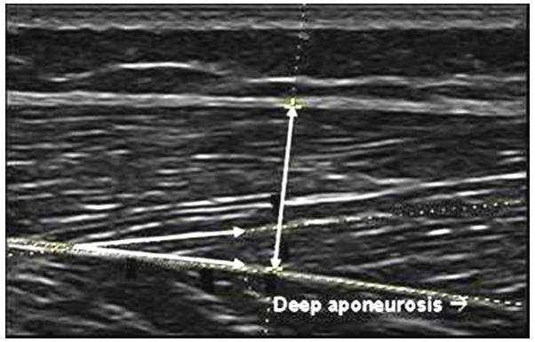
Figure 1.

Muscle thickness was calculated as the distance between superficial and deep aponeuroses in the middle of the ultrasound image at a 90° angle from the deep aponeurosis as indicated by the vertical line. Fascicle angle was the positive angle between the deep aponeurosis and the line of the fascicle as indicated by the intersecting lines.
