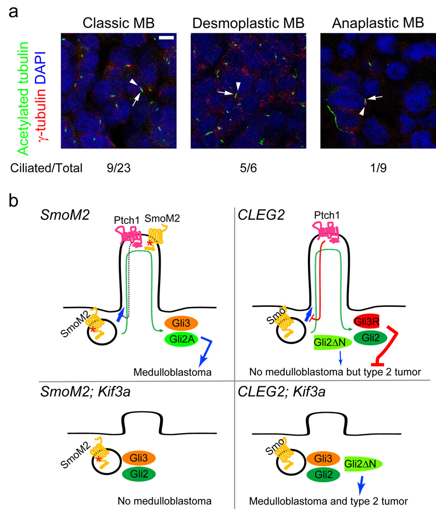
Fig. 3. Primary cilia are present in a subset of human medulloblastomas.
(a) Nine of twenty-three classic and five of six desmoplastic medulloblastomas have primary cilia in most cells. Eight of nine anaplastic medulloblastomas have few or no ciliated cells (shown is an example of non-ciliated anaplastic medulloblastoma). Cilia and basal bodies are stained by anti-acetylated tubulin (green, arrows) and anti-γ-tubulin (red, arrowheads), respectively. Scale bar = 5 µm. (b) Model summarizing results of dual roles of primary cilia in SmoM2- and Gli2ΔN-driven tumorigenesis. SmoM2 is insensitive to inhibition by Ptch1 and constitutively localizes to the cilia, where it activates Gli2 and inhibits Gli3 repressor (Gli3R) formation, leading to medulloblastoma (top left). Without the cilia, SmoM2 cannot activate downstream pathways; cerebellum remains small as in ciliary mutants8 and no tumors develop (bottom left). Gli2ΔN is constitutively active and independent of primary cilia, yet these mice do not develop medulloblastoma possibly due to thepresence of Gli3 repressor; however, these mice develop type 2 tumors later in life (top right). In the absence of cilia, Gli2ΔN induces medulloblastoma and type 2 tumors earlier in life. This may be due to the elimination of Gli3 repressor when cilia are removed (bottom right).

