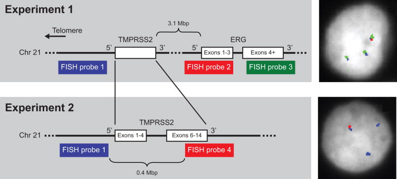
Figure 4.
Experimental designs to detect the presence of the TMPRSS2:ERG fusion by FISH. In Experiment 1, three probes were used to detect the fusion: a probe 5′ of TMPRSS2 (blue, probe 1), one encompassing the 5′ exons of ERG (red, probe 2), and one encompassing the 3′ exons of ERG (green, probe 3). In the normal configuration, the signals of all three probes overlap. The fusion is indicated by overlapping signals of probes 1 and 3 with loss or dissociation of probe 2. A CR tumor nucleus with two fusions and one normal probe configuration is shown to the right. In Experiment 2, we confirmed the presence of fusion with probe 1 and a second probe encompassing the 3′ exons of TMPRSS2 (red, probe 4) in adjacent tissue sections. Lone probe 1 signals indicate a deletion consistent with a TMPRSS2:ERG fusion. The panel to the right shows a positive nucleus from a section adjacent to the section used to capture the nucleus shown for Experiment 1. DAPI staining is indicated in gray; the hybridization signals are pseudocolored to correspond to the experimental schematics.

