
Figure 4.
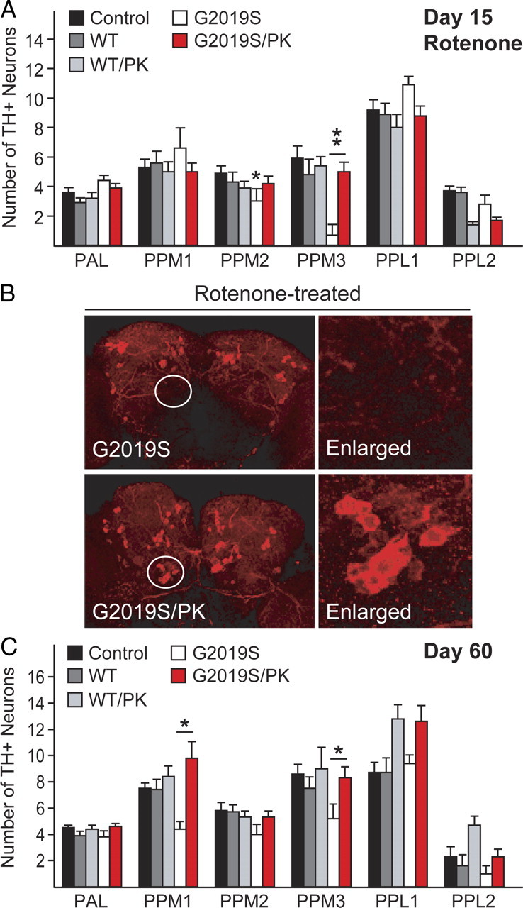
Parkin coexpression mitigates DA degeneration in LRRK2 G2019S-expressing flies in the presence or absence of rotenone. A, Bar graph showing the number of TH-positive DA neurons in different clusters of various fly species at 15 d after rotenone treatment (n = 15). B, Representative confocal microscopy images showing TH-positive (red) DA neurons in whole-mount adult brains derived from rotenone-treated flies expressing either LRRK2 G2019S alone or in the presence of parkin coexpression. Right, PPM3 cluster (circled) are shown at higher magnification. C, Bar graph showing the number of TH-positive DA neurons in different clusters of the various fly species at 60 d after eclosion, as indicated (n = 8) (genotype: ddc–Gal4/+ or ddc–Gal4–hLRRK2 or ddci-Gal4–hLRRK2;ddc–Gal4–hparkin).