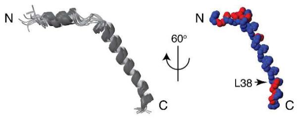
Figure 3.

Superimposition of 15 calculated backbone structures of Pf1 coat protein (left) and 60° rotation of one structure to the vertical axis (right). A slight kink at residue 38 is indicated. Distribution of hydrophilic (red) and hydrophobic (blue) residues demonstrates the amphipathic character of the short N-terminal in-plane helix and the C-terminal region.
