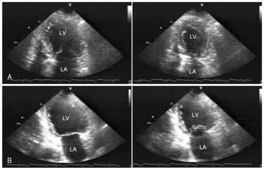
Fig. 3.
Echocardiography performed in December of 2003 showing decreased motion of the apical and mid-segments, with preserved contractility of the basal segments. A: apical 4 chamber view of end-diastole (Left) and end-systole (Right). B: apical 2 chamber view of end-diastole (Left) and end-systole (Right). LA: left atrium, LV: left ventricular.

