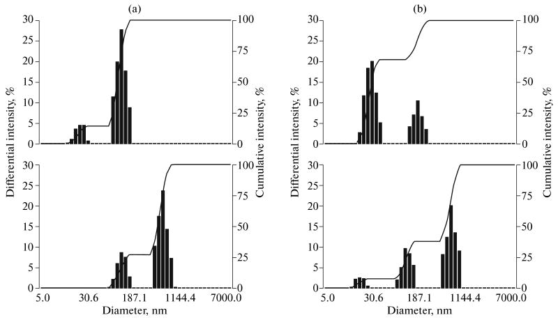
Fig. 2.
Particle size distribution: (a) empty micelles (0.25% PEG-PLLA solution) (at the top) and nanodroplets formed by 1 vol % PFP introduced into 0.25% PEG-PLLA solution (at the bottom, right peak); (b) paclitaxel-loaded micelles (0.25% PEG-PLLA solution) (at the top) and paclitaxel-loaded nanodroplets (at the bottom, right peak).

