
Fig. 5.
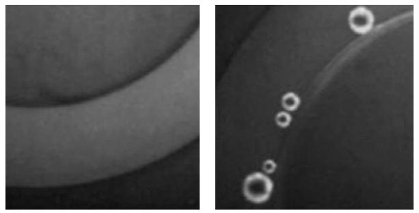
Localization of fluorescent DOX in microbubble walls (fluorescent microphotograph). On the left, there is the image of the microcapillary with a diameter of 340 μm (the light arc in the middle of the image) filled with the DOX-loaded nanoemulsion. The image is obtained at room temperature; the fluorescent nanodroplets are not resolved under the ×100 optical magnification, being maximal for the instrument setting used in the work. On the right, there are microbubbles formed under heating of the vessel, under vaporization of superheated nanodroplets and coalescence of formed vapor bubbles.