
Fig. 6.
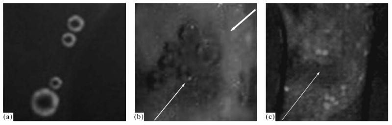
Fluorescence of microbubbles in the presence of the MBA MB231 breast cancer cells in the capillary with a diameter of 340 μm. Before the cell sonication, fluorescence of the microbubbles is observed; the cells do not exhibit fluorescence (a). After the sonication, the microbubble fluorescence decreases, whereas the cells, on the contrary, start fluorescing (b, c). Thus, under the action of ultrasound, DOX is released from the microbubbles and is internalized by the cells. The bubbles can be seen in some sites (b) and cannot be seen in the other, as in the image (c) that presents another site of the same sample. Survival of visible bubbles, although of lesser intensity (b), shows that the drug transfer from the bubbles into the cells does not require collapse of the bubbles. The figure is taken from [6].