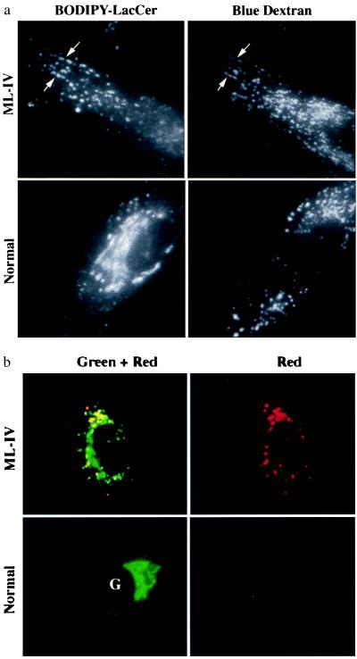
Figure 1.
Lysosomal accumulation of C5-DMB-LacCer in ML-IV cells. Normal and ML-IV fibroblasts were incubated overnight with a blue fluorescent dextran to label the lysosomes and subsequently pulse-labeled with C5-DMB-LacCer (see text). (a) In ML-IV cells, many of the punctate structures labeled with the fluorescent lipid colocalized with the dextran-stained lysosomes (e.g., at arrows), but in normal fibroblasts most of the fluorescent lipid was observed at the Golgi complex (G), with little present in the lysosomes. (b) Observations of BODIPY fluorescence in different regions of the spectrum also demonstrated a shift in BODIPY fluorescence toward red wavelengths in ML-IV (but not in normal) fibroblasts.

