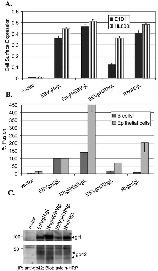
Figure 1. Rhesus gH is functional in EBV-mediated fusion with epithelial and B cells.
CHO-K1 cells were transiently transfected with EBV glycoproteins gp42 and gB and different combinations of gH and gL, as shown (Rh=rhesus). A. Postransfection, cells were transferred to 96 wells and assayed for gH/gL cell surface expression by CELISA with two different EBVgH/gL antibodies, E1D1 (black bars), a mouse monoclonal Ab, and HL800 (hatched bars), a rabbit polyclonal Ab. A representative experiment measured in triplicate is shown. B. Transfected CHO-K1 cells were overlaid with either Daudi B cells (black bars) or 293T kidney epithelial cells (gray bars) at 1:1 ratio and relative luciferase activity measured 24 hours later. Luciferase activity was normalized to wild-type levels, which was set to 100% for both cell types. Data are averages of three independent experiments with the standard deviations indicated by vertical lines. C. CHO-K1 cells were harvested 36 hours post transfection and the cell surface proteins labeled with biotin at 4°C. Biotinylated lysates were immunoprecipitated with the F-2-1 Ab (an anti-gp42 mouse monoclonal Ab) and probed with avidin-HRP in a Western blot. Proteins of interest are indicated with arrows. Positions of prestained protein markers (in kDa) are indicated.

