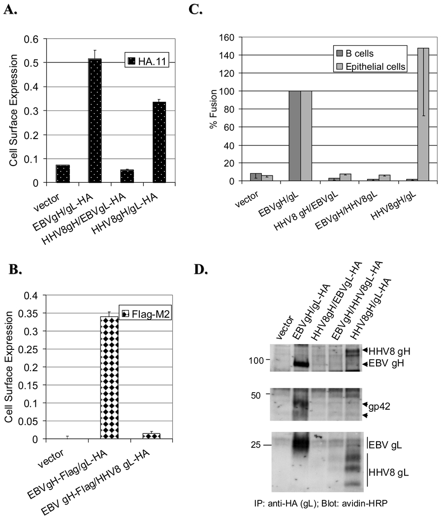
Figure 4. Heterologous complexes of EBV/HHV8 are not expressed at the cell surface and thus are nonfunctional in fusion.
As described above, CHO-K1 cells were transfected with EBV gp42, gB and different combinations of gH and gL, as indicated on the X-axes. A. The cell surface expression of different gH and gL complexes was assessed by CELISA as described before. Either an anti-HA (HA.11) (A) or anti-Flag (M2) antibody (B) was used. EBVgH construct with the Flag-tag was used to assess the cell surface expression of EBVgH/HHV8 gL complex since HHV8gL is expressed by itself at the cell surface (data not shown). No gp42 was included in transfection in B, since some EBVgH is expressed at the cell surface in the absence of EBVgL if gp42 is present (data not shown). Representative experiments performed in triplicate are shown. C. Posttransfection, CHO-K1 cells were overlaid with target cells and fusion assessed 24 hours later. Luciferase activity was normalized to wild-type levels, which was set to 100% for both cell types. Data are average of three independent experiments. D. Biotynylated lysates of transfected CHO-K1 cells were immunoprecipitated with an anti-HA Ab and Western probed with avidin-HRP. No association between HHV8 gH and/or gL with EBVgp42 was observed at the cell surface. The proteins of interest are indicated with arrows. Positions of prestained protein markers (in kDa) are indicated.

