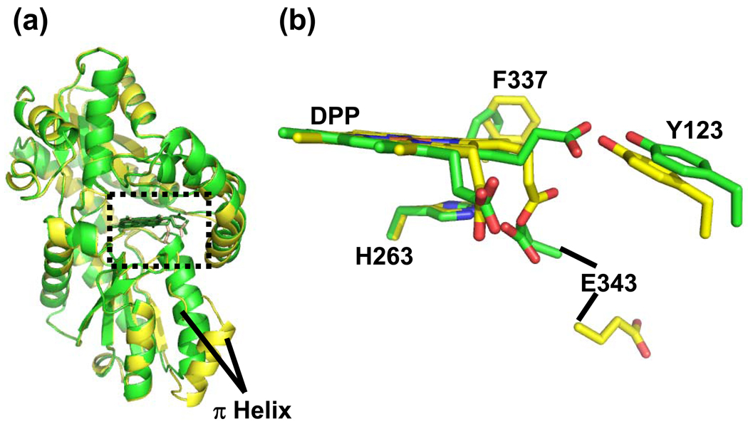
Figure 5.
Comparison of the MN-DEUT and NI-DEUT models. Panel a; Cartoon representation of secondary structure showing an overlay of the MN-DEUT (Green) and NI-DEUT (Yellow) models. The structurally conserved π helix is labeled and the active site is highlighted by a dashed box. Panel b; Model in stick format for the metallated deuteroporphyrin and the relative positions of some strictly conserved residues in both data sets. Carbon atoms in the MN-DEUT model are shown in green while the carbon atoms in the NI-DEUT model are shown in yellow. Oxygen, nitrogen, and metal (currently modeled as iron) atoms are colored red, blue and orange, respectively.

