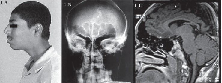
Figure 1.

(A) Photograph showing prominent superciliary arches, big nose and prognathism (B) Radiograph of skull showing prominent frontal and maxillary sinuses (C) MRI showing normal brain parenchyma and large sphenoid, frontal and ethmoidal sinuses with extensive pneumatization
