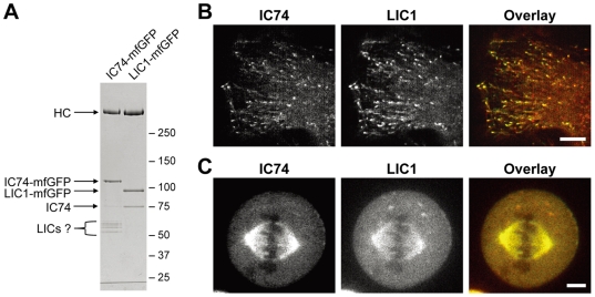
Figure 7. Fluorescent LIC1 as a probe for cytoplasmic dynein.
A. LIC1-mfGFP was purified from lysate of the LIC1-mfGFP expressing cells by StrepTrap chromatography and the protein composition was compared with that of the purified IC74-mfGFP fraction. Both heavy chain and IC74 were co-purified with LIC1-mfGFP. Polypeptides at 50–60 kDa range seen in the IC74-mfGFP fraction are undetectable in the purified LIC1-mfGFP fraction. B, C. IC74-mfGFP HeLa cells were transfected with LIC1-mCherry. Both the spot-like and the comet-like foci were labeled by LIC1-mCherry in interphase cells (B). Colocalization of IC74-mfGFP and LIC1-mCherry is also observed at mitotic spindle in metaphase cells (C). Scale bars: B, C, 10 µm.

