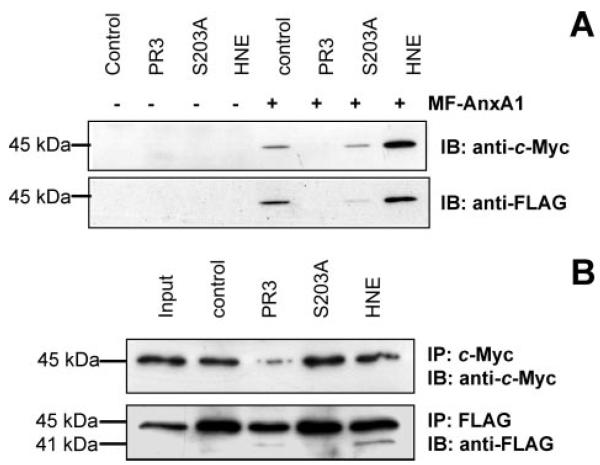
FIGURE 4. N-terminal proteolysis of MF-AnxA1 by PR3 in HMC1 cells.
A, HMC1 cells (control, PR3, PR3 mutant S203A, and HNE) were transfected with MF-AnxA1. After 24 h cell lysates were immunoblotted for c-Myc (upper panel) and FLAG (lower panel) with anti-c-Myc and anti-FLAG antibodies, respectively. B, extracts prepared from control, PR3, PR3 mutant S203A, and HNE cells were processed and incubated with MF-AnxA1 (input). Intact MF-AnxA1 (upper panel) and N-terminal-cleaved fragments (lower panel) were obtained as described above. Blots are representative from at least three distinct experiments.

