Figure 4.

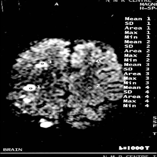
Diffusion-weighted image b = 1000 showing discrete hyperintense foci seen in right frontal white matter and in right frontoparietal region (study case images taken 9 h after ischemic stroke)

Diffusion-weighted image b = 1000 showing discrete hyperintense foci seen in right frontal white matter and in right frontoparietal region (study case images taken 9 h after ischemic stroke)