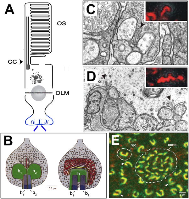
Fig. 5. The Synaptic Terminal.
A: Schematic of photoreceptor morphology; synaptic terminal is labeled in blue.
B: Schematic of a rod photoreceptor synapse. Views of the rod terminal perpendicular (left) and parallel (right) to the face of ribbon. Presynaptically, the ribbon tethers several hundred vesicles (white circles in red ribbon) and the active zone docks ca. 100 vesicles (yellow circles) and tethers ca. 770 vesicles. Postsynaptically, processes of 2 horizontal (h1, h2) and 2 bipolar (b1, b2) cells occupy the invagination. Reprinted with permission from (Rao-Mirotznik et al., 1995).
C. EM images of the “tetrad” ribbon synapse of a mammalian rod photoreceptor. Ribbon is indicated by an arrowhead and the inset is a confocal image of RIBEYE antibody staining. Reprinted with permission from (tom Dieck et al., 2005).
D. EM images of the “triad” ribbon synapse of a mammalian cone photoreceptor. Ribbons are indicated by arrowheads and the inset is a confocal image of RIBEYE antibody staining. Reprinted with permission from (tom Dieck et al., 2005).
E. Confocal image of a tangential section through macaque photoreceptor terminals. Dotted lines outline rod and cone presynaptic terminals immunostained for Bassoon (green), and the 1F subunit of an L-type Ca2C channel (red). The rod terminal typically comprises of a single, crescent-shaped ribbon, but can appear as two separate but linked ribbons (arrowheads). The cone terminal contains an array of smaller ribbons that serve separate invaginations. Reprinted with permission (Wassle, 2003).

