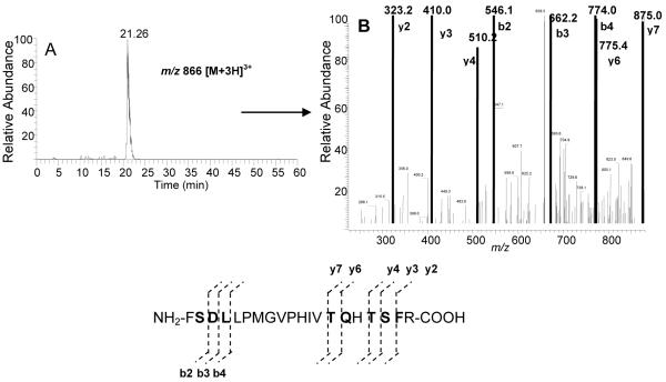
Figure 4.
ESI-LC-MS/MS analysis of the CYP 2B6 peptide spanning residues F359-R378 following inactivation by 17EE. (A) XIC of a triply charged peptide with a predicted m/z of 2596 (2284 + 312) (B) MS/MS analysis of the triply charged form of this peptide showed that the increase in mass occurred at the b2 ion or at S360 in the 2B6 sequence. Residues in bold were positively identified by MS/MS.

