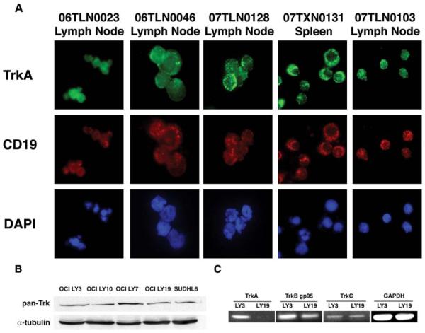
Figure 1.
B cells from primary Non-Hodgkin Lymphomas express TrkA. (A) A panel of single cell suspensions derived from patient biopsies of NHL were adhered to glass slides via cytospin. Immunofluorescence was performed using antibodies specific for CD19 (to identify B cells) and surface TrkA. Each patient’s cells expressed similar levels of TrkA on the surface of B cells derived from NHL tumors (TrkA: green, CD19: red and DAPI: blue). (B) DLBCL cell lines express Trk. The DLBCL cell lines were analyzed via immunoblot for expression of Trk receptors using a pan-Trk antibody, which recognizes TrkA, TrkB and TrkC. As shown, all cell lines exhibit pan-Trk expression. (C) The specific Trk receptors expressed was determined by RT-PCR in OCI-LY3 and OCI-LY19.

