
Figure 4.
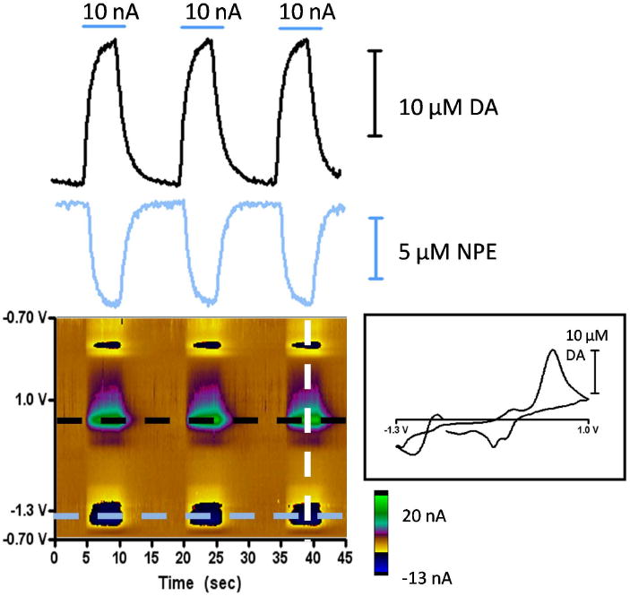
Analysis of consecutive iontophoretic ejections of dopamine and NPE. Carbon-fiber iontophoresis probes were used to eject a mixture of dopamine and NPE at pH 5.8 into a solution of TRIS buffer at pH 7.4. Upper left: The solid lines represent [DA] at 0.6 V (black) and [NPE] at -1.2 V (light blue) measured vs. time trace obtained for three consecutive 10 nA ejections at times indicated by the horizontal bars. Lower left: Color representation of the iontophoretic ejections shown, with applied voltage at the carbon fiber plotted vs. time and the current measured in response to dopamine and NPE ejection plotted in false color. Right: Cyclic voltammogram obtained at the time indicated by the white dashed line in the color plot.