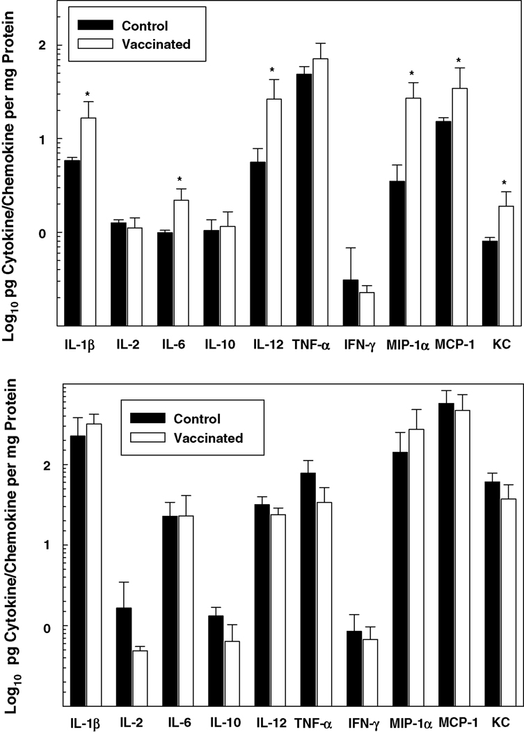
Figure 4. Proinflammatory cytokine and chemokine protein production is up-regulated in the lungs of vaccinated mice shortly following i.t. inoculation of F. tularensis LVS.
Groups of control and vaccinated mice were challenged i.t. with 6 LD50 F. tularensis LVS. At 4 hours (top panel) and 3 days (bottom panel) post-infection, the lungs were dissected, and the cytokines and chemokines listed were quantified by Bio-Plex cytokine bead array analysis. Data are the means ± SD derived from 4 mice treated comparably in each group. A second experiment yielded similar results. *Vaccinated group is significantly greater than control: P<0.05. Notably, IL-4 concentrations (not shown) were below the level of detection.

