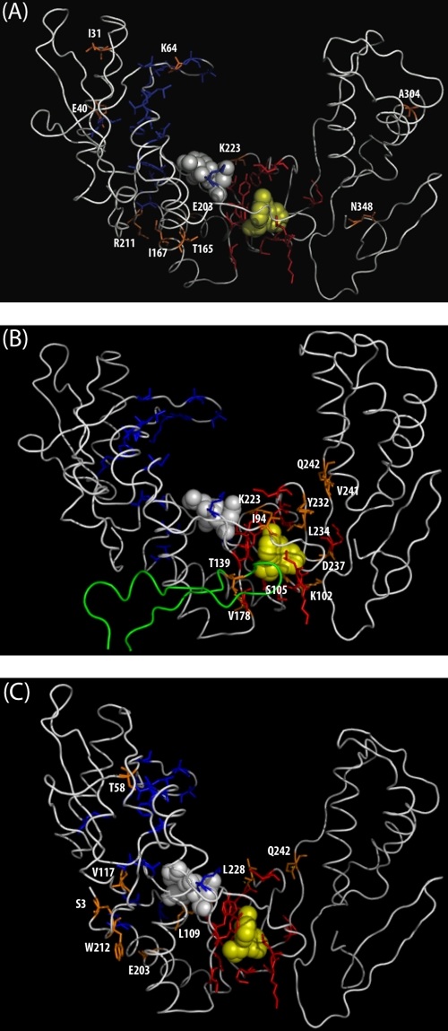
FIG. 2.
Three-dimensional structure (PDB accession number 3HVT) of HIV-1 RT bound to nevirapine showing residues 1 to 350 of the p66 monomer. The active-site residues D110, D185, and D186 are shown in space-fill mode in white. Nevirapine is shown in space-fill mode in yellow. Established NRTI resistance residues are shown in sticks mode in blue. Established NNRTI resistance residues are shown in sticks mode in red. Newly identified RTI-selected mutations are shown in sticks mode in orange. (A) Newly identified NRTI-selected mutations. (B) Newly identified NNRTI-selected mutations. Residues 120 to 148 in the p51 monomer are shown in green. (C) Newly identified undifferentiated mutations.

