FIG. 2.
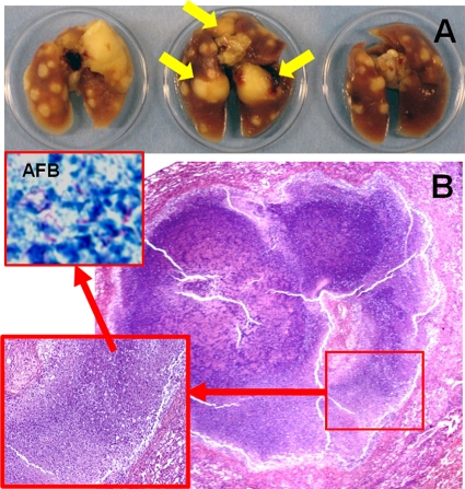
Mycobacterium tuberculosis-infected C3HeB/FeJ mice develop well-defined caseous granulomas. Unlike the BALB/c mouse model, C3HeB/FeJ mice display lung pathology with well-defined granulomas and central caseous necrosis after low-dose aerosol infection. (A) Formalin-fixed lungs 8 weeks after infection show caseous lesions. Yellow arrows point to the three large granulomas also visualized by [18F]FDG-PET imaging in the same animal at 6 weeks after infection (Fig. 3). (B) Lung histopathology from the same mice 8 weeks after infection, demonstrating well-defined granulomas with central necrosis and abundant acid-fast bacilli (AFB) (inset), is also shown.

