Abstract
A simple lateral-flow assay (LFA) based on a monoclonal antibody (MAb 70-17) was developed for the detection of foot-and-mouth disease virus (FMDV) under nonlaboratory conditions. The LFA was evaluated with epithelial suspensions (n = 704) prepared from current and historical field samples which had been submitted to the Pirbright Laboratory (United Kingdom) and from negative samples (n = 100) collected from naïve animals in Korea. Four FMDV serotypes (type O, A, Asia 1, and C) were detected in the LFA, but not the remaining three FMDV serotypes (SAT 1, SAT 2, and SAT 3). The diagnostic sensitivity of the LFA for FMDV types O, A, C, and Asia 1 was similar, at approximately 87.3%, to that of 87.7% obtained with antigen enzyme-linked immunosorbent assay (Ag-ELISA). The diagnostic specificity of the LFA was 98.8%, compared to 100% for the Ag-ELISA. These results demonstrate that the LFA using the FMDV MAb 70-17 to detect FMDV is a supportive method for taking rapid measurements at the site of a suspected foot-and-mouth disease outbreak in Asia before diagnosing the disease in the laboratory, thereby offering the possibility of implementing control procedures more rapidly.
The foot-and-mouth disease virus (FMDV), an Aphthovirus of the Picornaviridae family, causes a highly contagious and economically important disease in cloven-hoofed animals. Seven serotypes and many subserotypes have been identified. Foot-and-mouth disease (FMD) has been recognized as the most important constraint to international trade in animals and animal products (11, 12).
Typical cases of FMD are characterized by the formation of vesicles and epithelial erosions of the snout, tongue, hard and soft palate, coronary band, and feet. FMD cannot be distinguished clinically from other vesicular diseases, such as swine vesicular disease (SVD) and vesicular stomatitis (VS). Consequently, laboratory-based tests (virus isolation or demonstration of FMDV antigen or nucleic acid) are required for differential diagnosis. At present, routine diagnosis of FMD is made at several laboratories by the combined use of enzyme-linked immunosorbent assay (ELISA) and virus isolation techniques, supplemented by reverse transcriptase PCR (RT-PCR) (13-15, 18). However, most of these diagnostic methods require the availability of a dedicated laboratory facility, highly trained laboratory personnel, stable reagents, and multistep sample handling or preparation. In particular, virus isolation requires a laboratory cell culture facility, which can be difficult and expensive to maintain, besides requiring 4 to 6 days for test completion. Management of the logistical considerations associated with sample collection and transport is also required.
A rapid and easy-to-perform test, which would allow for on-site diagnosis to be made in the case of a suspected disease outbreak, would circumvent problems associated with the transportation of samples to the laboratory and would be especially useful for a faster diagnosis in areas where the disease is endemic. Since FMD is extremely contagious and long-distance airborne transmission is possible under favorable conditions, the need for and importance of rapid diagnosis of FMD have long been recognized. This was realized during the 2000 and 2002 FMD outbreaks in Korea, when the requirement for a pen-side diagnostic test that is quick and easy to perform, without the need for sophisticated equipment or laboratory expertise, was appreciated.
For these reasons, we have developed a rapid lateral-flow assay (LFA) based on FMDV antigen detection, which is easy to use and can be utilized on the farm to reduce the time required for transport and laboratory diagnosis. The detection of FMDV antigens by direct application of vesicular fluids and epithelial suspensions from animals of an infected farm may reduce the chances of diagnostic error arising from nonspecific reactions. The assay may be used to diagnose FMD at an early stage of infection and could be an effective tool in controlling FMD.
MATERIALS AND METHODS
Production of MAbs.
A number of monoclonal antibodies (MAbs) against FMDV were produced in BALB/c mice. Briefly, mice were immunized with an FMDV type O strain (SKR/O/2002). Conventional fusion and ELISA screening of resulting hybridomas were carried out. None of the resulting MAbs reacted with antigens of the SAT serotypes of FMDV, but three MAb clones (8-12, 17-9, and 70-17) were identified as exhibiting high specificity and reactivity against the other four FMDV serotypes (types O, A, Asia 1, and C) in ELISA and immunofluorescence assay, although they had no neutralizing ability against these viruses. Following preliminary tests, one MAb, designated MAb70-17 (isotype immunoglobulin G1), was chosen for incorporation into the LFA and for subsequent test validation, the results of which are currently presented.
Construction of lateral-flow device (LFD).
Gold particle suspensions (Fitzgerald Industries) were adjusted to pH 7.2 with 50 mM potassium carbonate (pH 9.6). MAb 70-17 was added dropwise to a 50-nm gold solution while being stirred to make a final concentration of 10 μg/ml, and the solution was kept stirring for 15 min. A 15% bovine serum albumin (BSA) solution was added at the rate of 30 μl per ml of gold particle suspension used. After being stirred for another 15 min, the coupled gold solution was centrifuged and the resulting supernatant was discarded in order to remove unbound MAb. Twelve milliliters of 2% BSA was added to the pellet of 200 ml of coupled gold solution, which was then sonicated in a sonic bath (Branson model 2200) in order to resuspend the pellet. The suspension was centrifuged again, and the final pellet was suspended in the same volume of 2% BSA (with 10 mM sodium carbonate, pH 9.6) and stored in a refrigerator at 4°C.
The MAb-coupled gold solution was diluted using dye dilution buffer (1% casein, 100 mM sodium phosphate, pH 7.0). The diluted gold solution was spread onto microglass paper (Lydall Inc.) and dried in a lyophilizer. Microglass paper was presoaked in pretreatment buffer (1% NP-40, 20 mM EDTA, 0.25% L-7600, 1% polyvinylpyrrolidone 10, 10 mM sodium phosphate, and 0.1% sodium azide, pH 7.0), dried on a fan after blotting of excessive liquid, and stored in a low-humidity room until use.
Cellulose filter paper (Millipore) was presoaked in pretreatment buffer (0.5% NP-40, 2% B-lactose, 1% polyethylene glycol 15000, 100 mM sodium phosphate, and 0.1% sodium azide, pH 7.0) and dried on a fan after blotting of excessive liquid. The prepared filter pad was stored in a low-humidity room.
An absorbent pad was attached along the long axis of the plate after the protective sheet from the tape at the top was peeled off. A dye pad was attached beneath the test membrane area along the long axis of the plate after the protective sheet from the tape at the bottom was peeled off. The dye pad overlapped the bottom of the test membrane by 2 to 3 mm. A filter pad then was attached to the plate to cover the bottom of the dye pad. The dressed membrane plate was cut into 0.665-cm-wide strips.
Test principle and procedure.
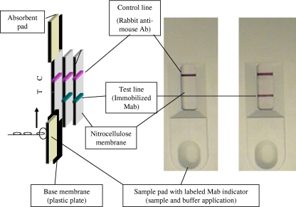
The LFA is a one-site immunometric assay using MAb 70-17 for the detection of FMDV antigen. MAb 70-17 to FMDV is conjugated with colored gold particles and is immobilized as a test line on a membrane. FMDV antigens present in the sample bind to the gold particles and form antigen-antibody-gold conjugate complexes, which migrate forward along the membrane. The complexes will be captured by the immobilized MAb 70-17, resulting in a colored band(s) in the test line. One or two bands are indicators of a positive test result. One band in the control line is significant for a negative test result, as the control antibody will bind the gold conjugate with both positive and negative samples and ensures correct test performance (Fig. 1).
FIG. 1.
Diagram of LFA. The clinical sample is added to the sample pad, where MAbs can interact with FMDV antigens. The addition of buffer enables the complex to migrate along the test strip, where gold-conjugated antibodies are captured by the immobilized MAbs at the test line. Excess material migrates further to be captured by anti-mouse antibodies at the control line to validate the procedure.
Test samples.
All serotype-inactivated FMDVs from a liquid-phase blocking ELISA kit (Pirbright Laboratory, United Kingdom) and two negative samples from Korea were used for preliminary evaluations. Epithelial suspensions (n = 704) were prepared from suspected cases of vesicular disease, using samples which had been submitted to the Pirbright Laboratory (United Kingdom) from 82 countries between 1992 and 2007. The samples consisted of different geographical origins and showed antigenic and molecular variations within each of the FMDV serotypes (n = 309), as well as others in which FMDV had not been detected (n = 389; classified as no virus detected [NVD]). Negative samples (n = 100) collected from naïve animals in Korea and samples from other vesicular diseases, i.e., SVD (n = 5) and VS (n = 1), were also tested.
Other tests.
All samples were tested by an indirect sandwich ELISA (Pirbright Laboratory, United Kingdom) to characterize the specificity of the virus serotype in original material and the cell culture antigens (6, 17). An optical density value of ≥0.1 above background indicated a positive reaction. If needed, a virus isolation test was also carried out using susceptible cell lines. The results of the ELISA were compared with those of LFA.
RESULTS
Characterization and selection of MAbs for use in LFD.
Eight hybridoma cell clones secreting anti-FMDV MAbs resulted from the fusion of mouse myeloma cells with spleen cells of a mouse immunized with an FMDV type O strain (SKR/O/2002). Three MAb clones (8-12, 17-9, and 70-17) had high reactivity against the FMDV antigen of serotypes A, Asia 1, O, and C in indirect ELISA and immunofluorescence assay but had no neutralizing ability against these serotypes (data not shown). Individual preliminary strip tests based on each of these three MAbs were performed, and the strip with MAb70-17 (isotype immunoglobulin G1) for both functions (capture and conjugated antibodies) was found to be the most appropriate for further test evaluation (data not shown).
Sensitivity and specificity of LFA.
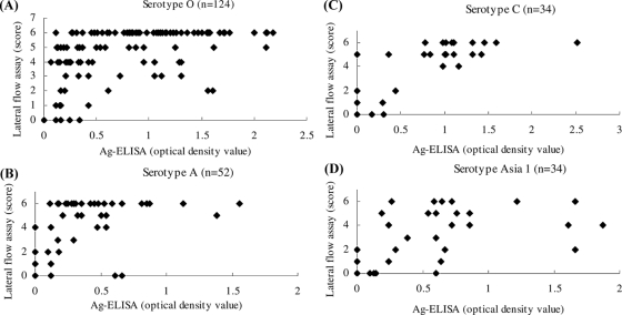
The LFA and antigen ELISA (Ag-ELISA) were performed with epithelial suspensions representative of each of the seven FMDV serotypes. The scoring results generated by the LFA were compared with optical density values derived from Ag-ELISA (Fig. 2). Results in the LFA were scored as follows: 0, negative reaction; 1, very weak reaction; 2, weak reaction; 3, weak to moderate reaction; 4, moderate reaction; 5, moderate to strong reaction; and 6, strong reaction. A mean optical density value of ≥0.1 in the ELISA was taken as indicative of a positive reaction. Although there was broad agreement between both methods for the detection of FMDV, a direct correlation between the results was not shown.
FIG. 2.
Comparison between reaction scores with the LFD and optical density values from ELISAs on different samples. (A) Serotype O; (B) serotype A; (C) serotype C; (D) serotype Asia 1. If the net optical density value in the ELISA was ≥0.1, the sample was considered positive. The scoring of the LFD was as follows: 0, negative; 1, very weak; 2, weak; 3, weak to moderate; 4, moderate; 5, moderate to strong; and 6, strong.
The results that were obtained with the LFA in comparison with those of the Ag-ELISA are summarized in Tables 1 and 2. The overall sensitivity and specificity of the LFA were similar, at approximately 86.9% and 98.8%, respectively, to those obtained by Ag-ELISA (87.7% and 100%, respectively). Both assays had similar sensitivities for each of the four FMDV serotypes O, A, Asia 1, and C. All but four samples positive by Ag-ELISA were also positive by LFA for type O. Four samples negative by LFA were borderline positive by Ag-ELISA. The sensitivity of the LFA was 93.5% when it was tested with type O samples (n = 124). For type A, 7 of 11 type A samples that were negative by Ag-ELISA were also scored negative by LFA, while the remaining 5 produced positive reactions in the LFA. The sensitivity of the LFA was 80% when tested with type A samples. One of four type Asia 1 samples that were negative by Ag-ELISA were also scored negative by LFA, while the remaining three produced a positive reaction in the LFA (two very weak and one weak) (n = 34). Four samples that were negative by LFA were positive by Ag-ELISA. Six of 11 type C samples that were negative by Ag-ELISA were also scored negative by LFA, while the remaining 5 produced positive reactions in the LFA. As shown in Table 1, all of the type SAT 1, SAT 2, and SAT 3 samples were negative by the LFA.
TABLE 1.
Sensitivity of LFA in comparison with FMDV Ag-ELISA
| Serotype | No. of samples tested | ELISA |
LFA |
||
|---|---|---|---|---|---|
| No. of positive samples | Sensitivity (%) | No. of positive samples | Sensitivity (%) | ||
| O | 124 | 120 | 96.8 | 116 | 93.5 |
| A | 52 | 41 | 78.8 | 42 | 80.7 |
| Asia 1 | 34 | 30 | 88.2 | 28 | 82.3 |
| C | 34 | 23 | 67.6 | 26 | 76.5 |
| SAT 1 | 24 | 12 | 50.0 | 0 | 0 |
| SAT2 | 35 | 30 | 85.7 | 0 | 0 |
| SAT3 | 6 | 5 | 83.3 | 0 | 0 |
| Totala | 244 | 214 | 87.7 | 212 | 86.9 |
Total numbers of samples testing positive and derived sensitivity values calculated for FMDV serotypes O, A, Asia 1, and C.
TABLE 2.
Specificity of LFA in comparison with FMDV Ag-ELISAa
| Sample group | No. of samples tested | LFA |
|
|---|---|---|---|
| No. of positive samples | Specificity (%) | ||
| SVDV | 5 | 0 | 100 |
| VSV | 1 | 0 | 100 |
| Negative epitheliab | 100 | 0 | 100 |
| NVD | 389 | 6 | 98.5 |
| Total | 495 | 6 | 98.8 |
For all samples tested, there were no positive ELISA results, giving a specificity of 100% for the Ag-ELISA.
From naïve animals.
Five samples of SVD virus (SVDV) and one sample of VS virus (VSV) were all negative in the LFA. One hundred negative samples from naïve animals were negative by Ag-ELISA and LFA. Only 6 of 389 NVD samples were positive by LFA, and the specificity of the LFA was 98.5%. The overall specificity with SVDV, VSV, negative, and NVD samples was 98.8%.
DISCUSSION
An essential component of any disease control strategy includes the deployment of diagnostic assays to rapidly confirm the initial clinical determination of infection. For FMD, this is of particular importance because FMDV is highly infectious and clinically indistinguishable from infection due to SVDV and VSV. At present, detection of FMDV is performed by a combination of Ag-ELISA, cultivation in cell cultures, and RT-PCR (5, 6, 13-15, 17). These methods are reliable and accurate but require skill and a complex laboratory and take several hours to perform. Transportation of clinical samples from the suspected outbreak sites to the laboratory also is necessary, takes time, and can be a major constraint in making rapid and accurate decisions for disease control. Researchers have been examining alternative assay systems that allow more rapid confirmation of clinical diagnosis but which do not require a laboratory setting and may be performed at the pen side instead (4, 8). Efforts have been made to develop portable real-time PCR machines with accessory kits. However, such methodology has yet to be validated fully and may be expensive (10).
During the devastating outbreak of FMD in Korea in 2002, there was a 24-h stamping-out policy, which meant that farm livestock of farms showing clinical signs conducive to FMD was condemned. However, accurate clinical diagnosis is important and can be fraught with problems, as there are a number of syndromes due to other causes that can easily be confused with FMD (2). There can be serious consequences of false-positive or false-negative results, and rapid, on-the-spot, reliable diagnostic tests to support clinical suspicions are required. The purpose of this study was to develop a supportive method for taking rapid measurements at the site of a suspected FMD outbreak before diagnosing the disease in the laboratory, thereby offering the possibility of implementing control procedures more rapidly.
The LFA is an appropriate technology on which to base a rapid assay. The technique permits rapid diagnosis, allowing time for the early implementation of control measures to reduce the possibility of spread of FMD. The LFA has been developed widely to support clinical diagnosis of different diseases (1, 3, 9), including FMD (7, 16).
At the commencement of this study, it was known that the MAb utilized for the particular FMD LFD reported by Reid et al. (16) was no longer viable, and as an alternative device was not yet available, the main purpose of this study was to identify a substitute MAb for test development. To meet this need, eight MAbs were evaluated for the ability to react with FMDVs of all seven serotypes and for their specificity of reaction against SVDV and VSV by ELISA. None of these reacted with FMDVs of the SAT FMDV serotypes, but three had a high degree of reaction against FMDVs of the O, A, C, and Asia 1 serotypes (the FMDV serotypes that historically have been associated with circulation in Asia), did not react with SVDV or VSV, and therefore warranted further investigation for incorporation into an LFA. Following preliminary investigations on prototype LFA devices, one MAb (MAb70-17) was selected for further investigation, with the results presented.
Although the LFA did not react with FMDVs of all serotypes, the sensitivity (86.9%) and specificity (98.8%) of the LFA are similar to those of Ag-ELISA for the detection of FMDV antigen. The Ag-ELISA is laborious and complicated and also needs serotype-specific polyclonal antibodies for operation; however, the LFA is more rapid and effective. The disagreement between both assays for the detection of FMDV may be due to either of the following causes. First, the majority of ELISA results at the World Reference Laboratory were derived at the time of sample receipt, while the companion LFD tests were performed later on suspensions that had subsequently been stored at −80°C and whose quality may have deteriorated in the interim. Second, differences in the affinities of the antibodies used in the two assays (serotype-specific polyclonal antibodies are used in the ELISA and a MAb is used in the LFD) may account for a difference in their intratypic spectra of reactions against field virus strains.
The results illustrate that a one-step LFA for the detection of FMDV antigens in specimens from animal vesicular and epithelial fluids has successfully been developed. Test results can be available within 20 min of collection of a sample from an animal. The LFD does not serotype the causative FMDV, but this is defined from any primary outbreak in a previous FMD area as a consequence of other laboratory-based test procedures. The usefulness of the device is anticipated from its deployment in secondary cases of disease when confirmation of FMD is all that may be required to allow appropriate control measures to be implemented. Since the LFA is simple and easy to use, it can be adopted in underdeveloped countries where diagnostic facilities are limited. It can serve as an alternative means of sample testing or be an addition to existing laboratory-based test procedures, help in the implementation of appropriate control measures, and reduce the amount of unnecessary animal culling.
Historically, FMDVs of only four serotypes (O, A, Asia 1, and C) have been identified in Asia, and consequently, this LFA would be a very useful aid in the diagnosis of FMD in all animal species which might become infected in outbreak situations in the region. However, since the introduction of FMDVs of the SAT serotypes to the region could conceivably occur, future efforts will concentrate on selecting a SAT group-specific MAb(s) as an extra component to counter the disadvantage of the failure of the LFD to detect these FMDVs and to make it useful for deployment in regions of the world where these FMDV types are prevalent.
Acknowledgments
We thank Nam-Kyu Shin and his colleagues for arrangement and collaboration on the project.
This project was supported by a grant from the National Veterinary Research and Quarantine Service (NVRQS), Republic of Korea, and partly by Princeton BioMeditech Corporation. The work of N.P.F. was supported financially by the Department for the Environment, Food and Rural Affairs (DEFRA; project numbers SE1120 and SE1123).
Footnotes
Published ahead of print on 2 September 2009.
REFERENCES
- 1.Al-Yousif, Y., J. Anderson, C. Chard-Berqstom, and S. Kapil.. 2002. Development, evaluation and application of lateral-flow immunoassay (immunochromatography) for detection of rotavirus in bovine fecal samples. Clin. Diagn. Lab. Immunol. 9:723-725. [DOI] [PMC free article] [PubMed] [Google Scholar]
- 2.Ayers, E., E. Cameron, R. Kemp, H. Leitch, A. Mollison, I. Muir, H. Reid, D. Smith, and J. Sproat. 2001. Oral lesions in sheep in Dumfries and Galloway. Vet. Rec. 148:720-723. [PubMed] [Google Scholar]
- 3.Brüning, A., K. Bellamy, D. Talbot, and J. Anderson. 1999. A rapid chromatographic strip test for the pen-side diagnosis of rinderpest virus. J. Virol. Methods 81:143-154. [DOI] [PubMed] [Google Scholar]
- 4.Callahan, J. D., F. Brown, F. A. Osorio, J. H. Sur, E. Kramer, G. W. Long, J. Lubroth, S. J. Ellis, K. S. Shoulars, K. L. Gaffney, D. L. Rock, and W. M. Nelson. 2002. Use of a portable real-time reverse transcriptase-polymerase chain reaction assay for the rapid detection of foot-and-mouth disease virus. J. Am. Vet. Med. Assoc. 220:1636-1642. [DOI] [PubMed] [Google Scholar]
- 5.Callens, M., and K. De Clercq. 1997. Differentiation of the seven serotypes of foot-and-mouth disease virus by reverse transcriptase polymerase chain reaction. J. Virol. Methods 67:35-44. [DOI] [PubMed] [Google Scholar]
- 6.Ferris, N. P., and M. Dawson. 1988. Routine application of enzyme-linked immunosorbent assay in comparison with complement fixation for the diagnosis of foot-and-mouth and swine vesicular diseases. Vet. Microbiol. 16:201-209. [DOI] [PubMed] [Google Scholar]
- 7.Ferris, N. P., A. Nordengrahn, G. H. Hutchings, S. M. Reid, D. P. King, K. Ebert, D. J. Paton, T. Kristersson, E. Brocchi, S. Grazioli, and M. Merza. 2009. Development and laboratory validation of a lateral flow device for the detection of foot-and-mouth disease virus in clinical samples. J. Virol. Methods 155:10-17. [DOI] [PubMed] [Google Scholar]
- 8.Hearps, A., Z. Zhang, and S. Alexandersen. 2002. Evaluation of the portable Cepheid SmartCycler real-time PCR machine for the rapid diagnosis of foot-and-mouth disease. Vet. Rec. 150:625-628. [DOI] [PubMed] [Google Scholar]
- 9.Kameyama, K., Y. Sakoda, K. Tamai, H. Igarashi, M. Tajima, T. Mochizuki, Y. Namba, and H. Kida. 2006. Development of an immunochromatographic test kit for rapid detection of bovine viral diarrhea virus antigen. J. Virol. Methods 138:140-146. [DOI] [PubMed] [Google Scholar]
- 10.King, D. P., J. P. Dukes, S. M. Reid, K. Ebert, A. E. Shaw, C. E. Mills, L. Boswell, and N. P. Ferris. 2008. Prospects for rapid diagnosis of foot-and-mouth disease in the field using reverse transcriptase-PCR. Vet. Rec. 162:315-316. [DOI] [PubMed] [Google Scholar]
- 11.Kitching, R. P. 1999. Foot-and-mouth disease: current world situation. Vaccine 17:1772-1774. [DOI] [PubMed] [Google Scholar]
- 12.Leforban, Y. 1999. Prevention measures against foot-and-mouth disease in Europe in recent years. Vaccine 17:1755-1759. [DOI] [PubMed] [Google Scholar]
- 13.Reid, S. M., M. A. Forsyth, G. H. Hutchings, and N. P. Ferris. 1998. Comparison of reverse transcription polymerase chain reaction, enzyme linked immunosorbent assay and virus isolation for the routine diagnosis of foot-and-mouth disease. J. Virol. Methods 70:213-217. [DOI] [PubMed] [Google Scholar]
- 14.Reid, S. M., G. H. Hutchings, N. P. Ferris, and K. De Clercq. 1999. Diagnosis of foot-and-mouth disease by RT-PCR: evaluation of primers for serotypic characterisation of viral RNA in clinical samples. J. Virol. Methods 83:113-123. [DOI] [PubMed] [Google Scholar]
- 15.Reid, S. M., N. P. Ferris, G. H. Hutchings, A. R. Samuel, and N. J. Knowles. 2000. Primary diagnosis of foot-and-mouth disease by reverse transcription polymerase chain reaction. J. Virol. Methods 89:167-176. [DOI] [PubMed] [Google Scholar]
- 16.Reid, S. M., N. P. Ferris, A. Brüning, G. H. Hutchings, Z. Kowalska, and L. Åkerbolm. 2001. Development of a rapid chromatographic strip test for the pen-side detection of foot-and-mouth disease virus antigen. J. Virol. Methods 96:189-202. [DOI] [PubMed] [Google Scholar]
- 17.Roeder, P. L., and P. M. Blanc Smith. 1987. Detection and typing of foot-and-mouth disease virus by enzyme-linked immunosorbent assay: a sensitive, rapid and reliable technique for primary diagnosis. Res. Vet. Sci. 43:225-232. [PubMed] [Google Scholar]
- 18.Vangrysperre, W., and K. De Clercq. 1996. Rapid and sensitive polymerase chain reaction based detection and typing of foot-and-mouth disease virus in clinical samples and cell culture isolates, combined with a simultaneous differentiation with other genomically and/or symptomatically related viruses. Arch. Virol. 141:331-344. [DOI] [PubMed] [Google Scholar]


