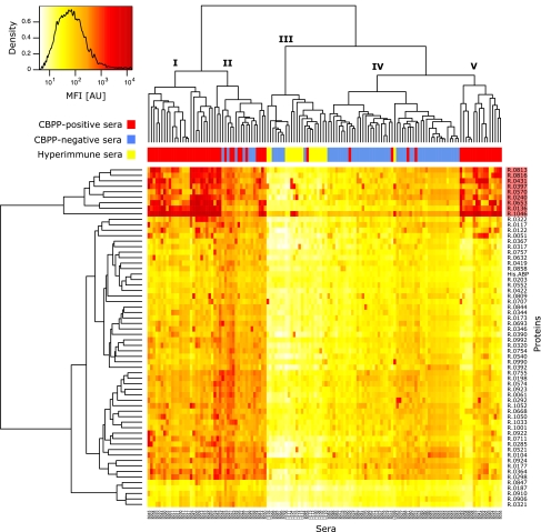
FIG. 1.
Cluster analysis of screening data. Heat map overview of log10-transformed MFI data from the bead-based screening of 134 samples (115 sera) against 61 recombinant M. mycoides SC surface proteins and His6ABP. Color intensity denotes signal intensity. Proteins against which large amounts of serum antibodies were detected in CBPP-positive sera, thus suggesting diagnostic importance, formed a cluster of nine proteins (top of vertical dendrogram; the cluster is highlighted in red). Horizontally, sera were separated into five groups. Clusters I, II, and V contained a majority of the CBPP-positive samples (red). Cluster III contained all but one hyperimmune serum (yellow), which displayed signals of low intensities, while cluster IV contained mostly CBPP-negative bovine sera (blue) as well as the positive control antiserum to M. mycoides SC PG1. All technical replicates clustered together, except for one specimen of a Swedish negative control serum.

