FIG. 3.
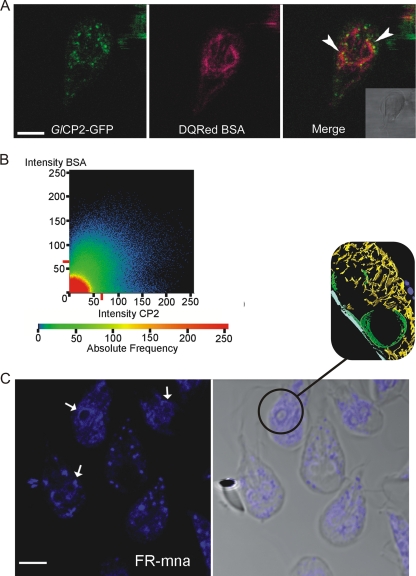
Giardia orthologues of mammalian lysosomal proteases are present in the endocytic TVN, where they colocalize with endocytosed proteins. (A) The cathepsin B-like protease GlCP2::GFP reporter (green, Giardia orthologue of mammalian lysosomal proteases) colocalized with endocytosed albumin (red, DQ Red BSA) during live cell imaging. Yellow in the merged image shows overlap (arrowheads) in both the perinuclear region of the trophozoite and the finer network of the TVN. Validation of this overlap is shown in panel B. Bar, 5 μm. (B) Quantification of colocalization shown by plotting the pixel fluorescence intensities from channels for DQ Red BSA and GlCP2::GFP. The majority of pixels are positive for both markers. (C) Cysteine protease activity detected by fluorescent substrate cleavage (Z-FR-MNA). This activity is seen in the outer TVN but is particularly intense in the perinuclear region (arrows). The inset shows perinuclear cisternae and TVN from tomography in Fig. 1 as a correlate. Scale bar, 5 μm.

