FIG. 6.
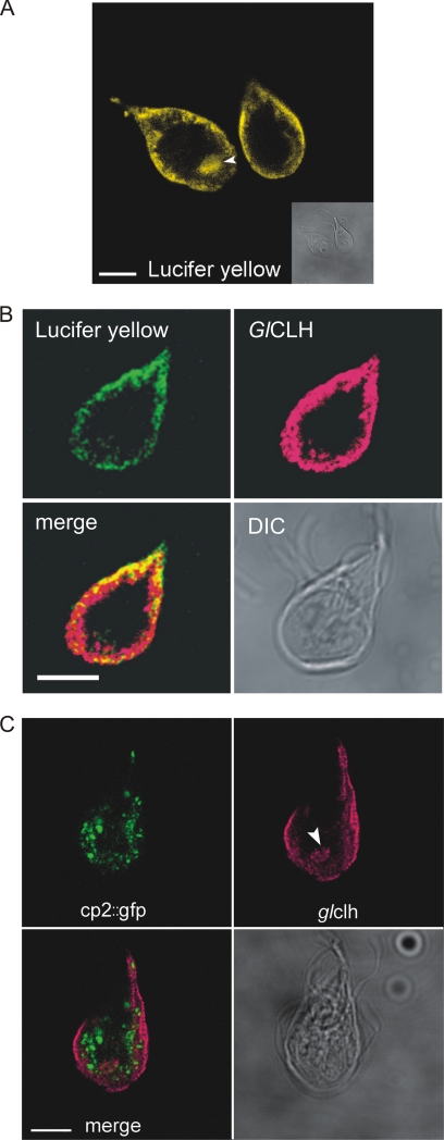
Cathepsin-like cysteine proteases are not concentrated in the acidified clathrin-rich PVs of Giardia trophozoites. (A) Lucifer yellow staining (yellow) highlights the acidic nature of the PV network aside from the cell periphery. PVs are also abundant in an area of the cell near the ventral flagellar exit site (arrowhead). Scale bar, 5 μm. (B) Clathrin (red) and Lucifer yellow (green) overlap at the PV network. Colocalized pixels appear yellow on the merged image. Scale bar, 5 μm. (C) Cathepsin B-like cysteine protease GlCP2::GFP (green) marks the endocytic TVN of Giardia. This network is distinct from the clathrin-associated PVs (red). Occasional colocalization of GlCP2::GFP with clathrin was seen at the interface between the two staining patterns but was the exception. Clathrin is also abundant near the exit site of the ventral flagella (arrowhead). Scale bar, 5 μm.

