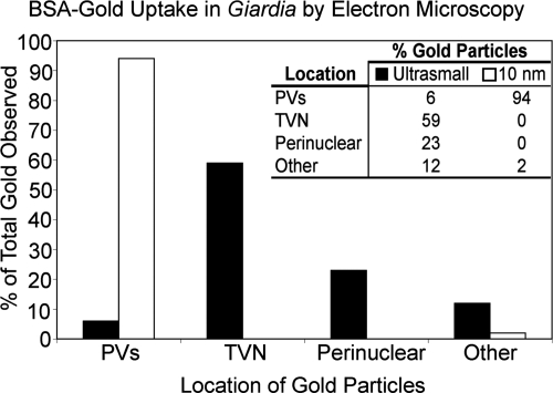
FIG. 7.
Ultrastructural analysis revealed that gold particles were endocytosed into the TVN. By EM, endocytosed albumin-coated ultrasmall (0.8-nm) gold particles localized mainly to the tubular TVN and the perinuclear region. This correlated with the endocytic pattern seen in the TVN and perinuclear region by confocal analysis in Fig. 1 to 3. Endocytosis of larger (10-nm) albumin-coated gold particles was arrested in the PVs. At least three electron microscopy images were analyzed for each gold particle size.

