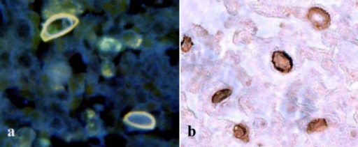
FIG. 1.
Localization of cellulose in the Acanthamoeba cyst wall after D-CBD staining with two different conjugates of frozen sections of corneas from a patient with keratitis. (a) Note the distinct binding to the inner cyst wall and the orange autofluorescence of the outer cyst wall after staining with the D-CBD-Alexa Fluor 350 blue conjugate. There is some blue autofluorescence of the connective tissue background. (b) Identification of Acanthamoeba cyst in corneal tissue stained with biotinylated D-CBD.

