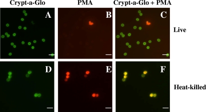
FIG. 1.
Microscopic analyses distinguishing live from dead C. parvum oocysts using PMA. Live (A to C) or heat-killed (D to F) (70°C, 20 min) C. parvum oocysts treated with PMA and stained with Crypt-a-Glo. Crypt-a-Glo-labeled oocysts (green) are shown in panels A and D; the same cysts stained with PMA (red) are shown in panels B and E; and panels C and F represent overlaid images with Crypt-a-Glo and PMA (yellow) staining. Bar = 10 μm.

