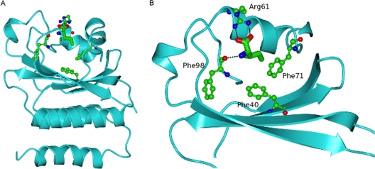
FIG. 1.
Ribbon diagram of the GAF domain of B. subtilis CodY with the bound isoleucine ligand (cylinder representation) surrounded by residues whose side chains have been mutated in this study. (A) The whole GAF domain is shown. (B) Close-up of the ligand-binding pocket in which the lower helical region has been omitted and residues 70 to 96 have been left out for clarity of view. Polar interactions with the isoleucine ligand are indicated by dashed lines.

