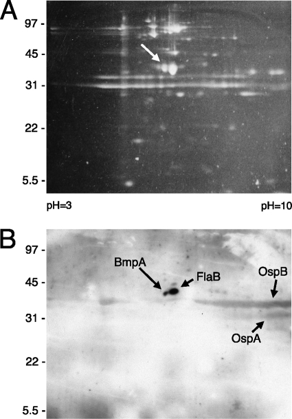
FIG. 1.
Two-dimensional electrophoretic analysis of an outer membrane-enriched fraction of cultured B. burgdorferi B31-MI-16. (A) Polyacrylamide gel stained with the fluorescent dye SYPRO Ruby. The arrow indicates the protein spot that was determined to correspond with BmpA. (B) A second gel was run simultaneously to that shown in panel A; proteins were transferred to a nitrocellulose membrane and then examined for laminin-binding activities through ligand affinity blot analysis. Strong signals that were determined to correspond with BmpA and FlaB are indicated. Relatively weaker signals were produced from OspA and OspB, both of which often form broad smears across 2-dimensional gels (47).

