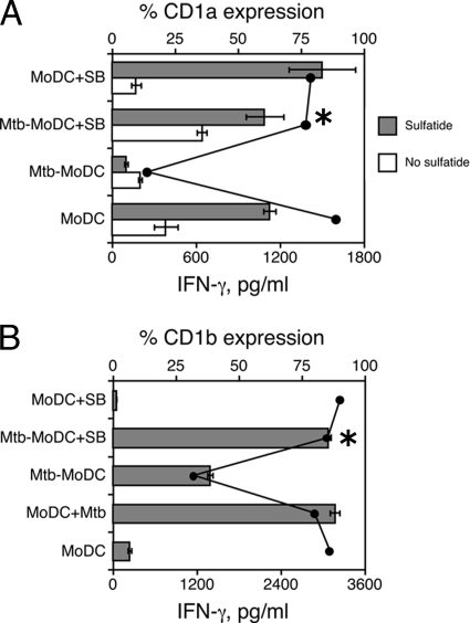
FIG. 3.
Inhibition of p38 restores the capacity of Mtb-MoDC to present lipid antigens to CD1-restricted T cells. The APC function of Mtb-MoDC was compared to that of control MoDC, DC derived from uninfected monocytes pretreated with the p38 inhibitor SB203580 (MoDC+SB), or M. tuberculosis-infected monocytes pretreated with the p38 inhibitor SB203580 (Mtb-MoDC+SB). In some experiments MoDC were infected with M. tuberculosis (MoDC+Mtb) at the end of the differentiation culture (day 5). (A) APC pulsed or not pulsed with sulfatide were cocultured with a sulfatide-specific CD1a-restricted T-cell clone. (B) A CD1b-restricted T-cell clone specific for Ac2SGL was used to test the capacity of infected cells to process mycobacteria and present lipid antigens. The bars indicate the amounts of IFN-γ secreted by responder T-cell clones, and the lines indicate the percentages of CD1 molecule expression on APC. The amounts of IFN-γ (mean ± standard deviation of three replicates) were measured after 48 h of culture. The results of one experiment that is representative of three experiments are shown. An asterisk indicates that there is a significant difference in IFN-γ secretion (P < 0.05) between M. tuberculosis-infected cells treated with SB203580 and nontreated cells.

