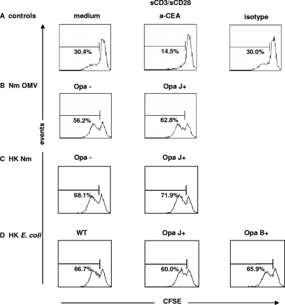
FIG. 6.
CD4+ T-cell responses to Opa proteins presented on OMV, heat-inactivated N. meningitidis, or a heterologous background. CD4+ T cells were prestimulated for 4 days with IL-2, labeled with CFSE, and then assessed for the ability to proliferate for 3 days in the presence of CD3/CD28. Additional agents included anti-CEACAM1 antibody A0115 or its isotype control (A), N. meningitidis strain H44/76 Opa− or OpaJ+ OMV (Nm OMV) (B), heat-killed N. meningitidis strain H44/76 Opa− or OpaJ+ (HK Nm) (C), and heat-killed wild-type OpaJ+ or OpaB+ E. coli (HK E. coli) (D). The number within each histogram represents the % divided cells in response to CD3/CD28, as measured by CFSE dilution (data are means of triplicate samples representative of three independent experiments).

