FIG. 1.
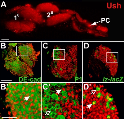
Differential Ush expression in the third-instar larval lymph gland. (A) Ush expression in primary and secondary lymph gland lobes (fluorescence microscopy) (magnification, ×20). (B to D′) Ush coexpression with hemocyte-specific markers in the primary lymph gland lobe (confocal microscopy) (magnification, ×60). (B and B′) Ush and the medullary zone prohemocyte marker DE-cad. (C and C′) Ush and the plasmatocyte marker P1 (Nimrod). (D and D′) Ush and the crystal cell marker lz-lacZ. Closed arrows indicate coexpression, and open arrows indicate a lack of coexpression. PC, pericardial cells. Scale bars, 50 μm (A and B) and 15 μm (B′).

