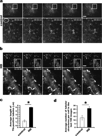
FIG. 5.
Long tubular intermediates formed in living HD fibroblasts viewed by TIRF. (a) Short tubular and small vesicular intermediates in control fibroblasts. White arrows point to a short tubule emerging and moving, whereas the oval surrounds two vesicles that fused with the plasma membrane and disappeared in the next image. The low signal is due to the small size of transferrin (Tfn)-positive structures. (b) Long tubules serve as transport intermediates in HD fibroblasts. Images are shown 4 s apart in order to show the change of tubular intermediates. Open arrows point to a tubule gradually fusing with the plasma membrane and becoming shorter and shorter, while the solid arrow indicates a growing tubule that becomes longer and longer. Scale bars, 10 μm. (c) Increase in the mean average length of tubules in HD fibroblasts ± SD (n = 72 tubules from five control cells and 72 tubules from five HD cells; *, P < 0.001, determined by the Student t test). (d) Quantification of the mean number of tubular intermediates per cell observed in the last 100 images from each cell ± standard error of the mean (10 control and 10 HD cells) imaged by TIRF microscopy (*, P < 0.05, determined by paired Student t test).

