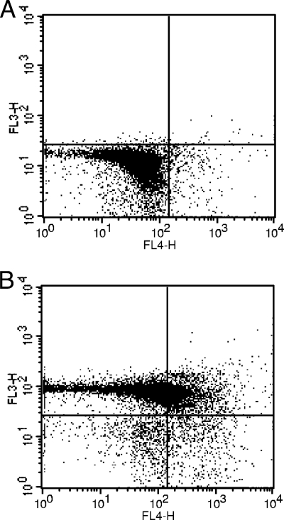
FIG. 5.
Invasion by isolate 4 induces apoptosis in porcine blood. Staining with annexin V-APC (FL4) and 7-AAD (FL3) determined phospholipid phosphatidylserine exposure and membrane integrity, respectively. (A) Two hours after the beginning of phagocytosis; (B) 6 h after the beginning of phagocytosis. When apoptosis is measured over time, cells can often be tracked from annexin V-APC and 7-AAD negative (viable, or no measurable apoptosis) (lower left) to annexin V-APC positive and 7-AAD negative (early apoptosis, membrane integrity present) (lower right) and finally to annexin V-APC and 7-AAD positive (end stage apoptosis and death) (upper right) (BD Biosciences).

