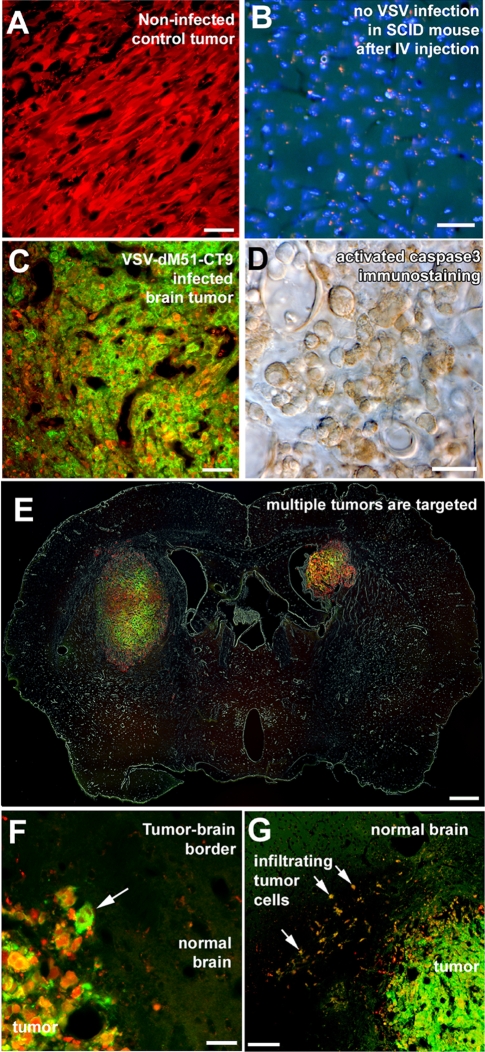
FIG. 6.
Neuroattenuated VSV-CT9-M51 targets human glioblastoma transplanted to mouse brains after a single intravenous injection. (A) Human brain tumor cells stably transfected with a red fluorescent protein grew with no signs of spontaneous regression when injected into SCID mouse brains. (B and C) Although an intravenous (IV) injection into control SCID mice did not result in any VSV infection in the brain (B), marked infection was found inside grafted tumors 72 h after intravenous injection of VSV-CT9-M51 (C). (D) Infection of the tumor resulted in widespread apoptosis, as documented by activated caspase 3 immunostaining. (E) Multifocal tumors within the brain were simultaneously infected after intravenous injections. (E and F) Despite robust VSV-CT9-M51 infection inside the tumor (arrow in panel F), no infection was observed outside the tumor anywhere in the mouse brain. (G) Tumor cells that grew infiltratively (arrows), away from the main tumor bulk into the normal brain parenchyma, were also infected, along with the main tumor. Scale bars, 50 μm (A to C), 20 μm (D), 500 μm (E), 25 μm (F), and 300 μm (G).

