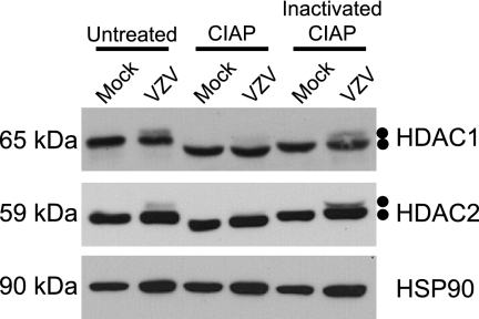
FIG. 2.
Analysis of HDAC1 and HDAC2 in infected-cell extracts following incubation with CIAP. MeWo cells were mock infected or infected with cell-associated wild-type VZV at a multiplicity of infection of 0.025 and harvested at 72 hpi. At 72 hpi, all of the cells were infected as determined by phase microscopy and/or ORF66p-GFP expression. Lysates from mock- and VZV-infected cells were reacted with dephosphorylation buffer alone (untreated), CIAP, or inactivated CIAP as described in Materials and Methods. Equal amounts of total protein (20 μg) from each sample were analyzed by Western blotting using antisera specific for HDAC1, HDAC2, and HSP90.

