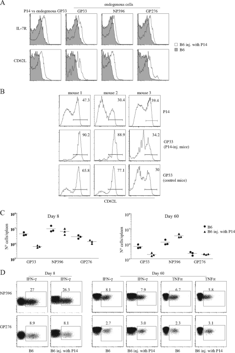
FIG. 4.
Impact of high-dose naïve Tg transfers on the endogenous response. B6.Ly5.1 mice left untreated or were injected with 5 × 10⁵ P14 Tg cells (Ly5.2+) and were infected simultaneously with 2 × 10⁵ PFU of LCMV Armstrong and studied at days 8 and 60 after infection. (A) CD62L and IL-7R expression in cells of with different peptide specificities at day 8 after infection. Histograms compare CD62L and IL-7R expression levels of CD8 cells with the indicated peptide specificities in 1 P14 injected (inj.) (open graphs) and 1 noninjected B6 mouse (gray) of 12 mice studied in two independent experiments. On the far left, P14 cells (open histogram) are compared to GP33-specific noninjected mice. (B) Variation of CD62L expression in GP33-specific cells 2 months after infection. Graphs compare Tg cells (upper) to endogenous cells present in the same mouse (middle). The lower graphs show endogenous cells in the mice that were not injected with P14 cells. (C) Absolute numbers of cells of different peptide specificity at day 8 (left) and 2 months (right) after infection. Results show individual mice from one experiment out of two with equivalent results. (D) IFN-γ expression after in vitro stimulation with NP396 and GP276 peptides at day 8 (left) and 2 months (right) after infection.

