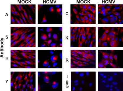
FIG. 1.
Immunofluorescence analysis of BiP shows that it is localized in two distinct locations during HCMV infection. Mock- or HCMV-infected cells were prepared for immunofluorescence microscopy at 96 hpi. BiP (red) was detected using the following antibodies described in Materials and Methods and Table 1: BiPA (A), BiPH (H), BiPR (R), BiPS (S), BiPY (Y), BiPC (C), and KDEL (K). Normal rabbit IgG (IgG) was used as a control. Nuclei were stained with DAPI (blue).

