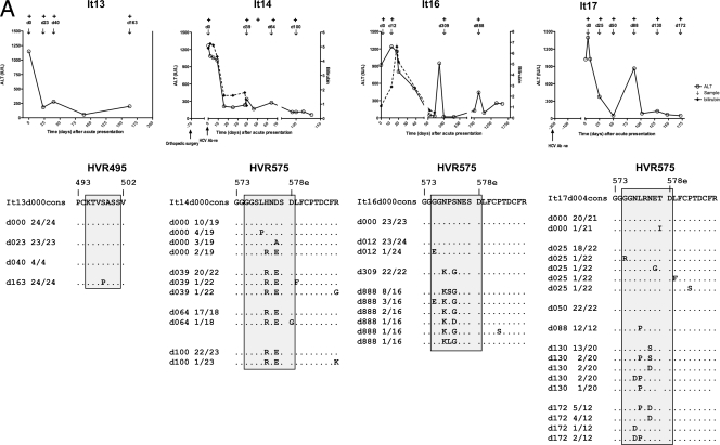
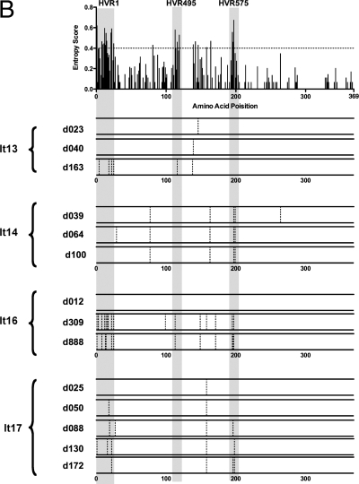
FIG. 6.
Evolution of HVR1 and HVR495 and HVR575 during primary HCV infection. (A) The clinical courses of four patients (It13, It14, It16, and It17) with an acute hepatitis secondary to primary HCV infection are shown. HCV RNA was detected by PCR at the time points shown (+). Patients were sampled at multiple time points when HCV RNA positive as indicated by “↓”. E2 quasispecies analysis was performed at each time point. HVR495 or HVR575 are shaded. The amino acid sequence of each variant is shown relative to the consensus sequence at the earliest time point (day 0). A dot indicates that the amino acid of the variant is identical to the consensus amino acid at day 0. The proportion of each variant/total number of variants is given. (B) The upper panel maps the position of the HVRs (shaded regions) derived from 18 patients with chronic HCV subtype 3a infection to the panels below. The data from four patients (It13, It14, It16, and It17) with acute infection are shown in the lower panels. These show mutations (represented by a dotted vertical bar) in the consensus sequence at each time point studied, relative to the consensus sequence at the earliest time point (day 0).


