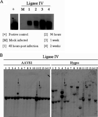
FIG. 6.
(A) Time course of junction formation in ligase IV hypomorphic cell lines. Cells were infected with wild-type AAV 2 at 105 viral particles/cell. Genomic DNA was isolated at the indicated time points for junction assay. (B) Southern hybridization of ligase IV clones infected with single-stranded AAV vectors P5PGKHygroGFP and SVAV2 (50:1 ratio, 105 vg/cell). Representative blots are shown. The left blots show signals for AAVS1 sequences, and the right blots show signals for hygromycin. The arrows indicate the clones that have both AAVS1 and hygromycin signals colinked.

