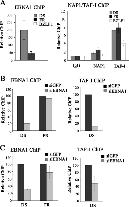
FIG. 6.
Localization of NAP1 and TAF-I to oriP elements by ChIP. (A) ChIP experiments were performed with Raji cells, using antibodies against EBNA1 (left), NAP1, TAF-I, or nonspecific rabbit IgG (right). Recovered DNA fragments were quantified by real-time PCR, using primer sets for the oriP DS and FR elements and the BZLF1 promoter region. The amplification signals were normalized to those from the same cell lysates prior to IP, using the same primer pairs. Signals from NAP1 and TAF-I antibody samples were expressed relative to that for the control IgG samples, which was set to 1. The results shown are from three independent experiments, with PCR quantification performed in triplicate for each experiment. (B and C) D98/Raji (B) and AGS-rEBV (C) cells were treated with siRNA against EBNA1 or GFP, and ChIP assays were performed for EBNA1 (left) and TAF-I (right) as in panel A.

