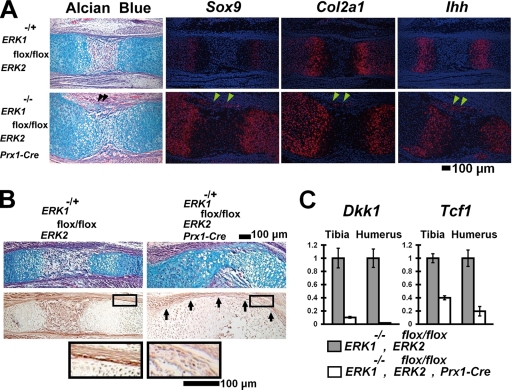
FIG. 4.
Ectopic cartilage formation in the perichondria of ERK1−/−; ERK2flox/flox; Prx1-Cre embryos. (A) Alcian blue staining and in situ hybridization of the femur at E15.5. The ectopic cartilage (arrowheads) in the perichondrium expresses Sox9, Col2a1, and Indian hedgehog (Ihh). (B) Alcian blue staining (top panel) and immunohistochemical staining of the radius for β-catenin (middle panel). ERK1−/−; ERK2flox/flox; Prx1-Cre embryos showed reduced β-catenin protein levels in the perichondrium at E16.5 (arrows). The boxed area is magnified in the bottom panel. (C) Tcf1 and Dkk1 expression quantitated by real-time PCR. ERK1−/−; ERK2flox/flox; Prx1-Cre embryos showed reduced Tcf1 and Dkk1 expression in the tibia and humerus at E16.5.

