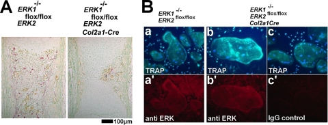
FIG. 8.
(A) TRAP staining of the femur showing decreased TRAP-positive cells in ERK1−/−; ERK2flox/flox; Col2a1-Cre embryos at E16.5. (B) ELF97-based fluorescent TRAP staining in combination with immunofluorescence for ERK protein. Spleen cells from ERK1−/−; ERK2flox/flox (a) and ERK1−/−; ERK2flox/flox; Col2a1-Cre (b,c) embryos formed TRAP-positive, multinucleated osteoclast-like cells in the presence of M-CSF and RANKL. Nuclei were visualized by DAPI (4′,6-diamidino-2-phenylindole). Lower panels show immunofluorescence using anti-ERK antibody (a′ and b′) or nonimmune immunoglobulin G (IgG) (c′) in corresponding cells.

