FIG. 9.
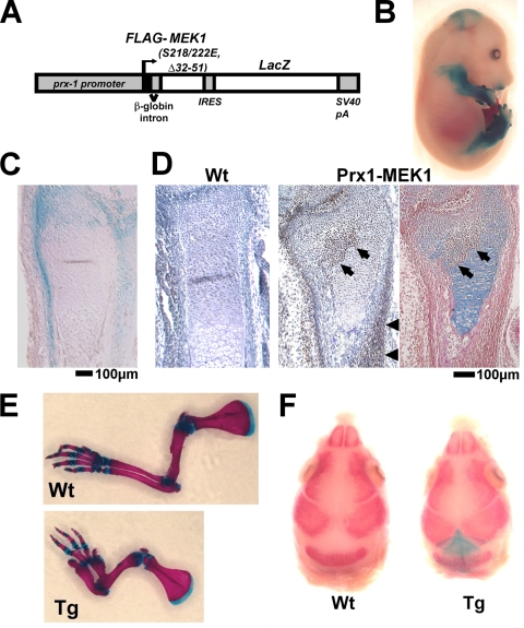
(A) Schematic representation of the construct that drives the expression of a constitutively active mutant of MEK1 and LacZ under the control of a 2.4-kb prx1 promoter. (B) X-Gal staining of an E15.5 embryo showing transgene expression in the limb and cranium. (C) X-Gal staining of the distal ulna of an E15.5 embryo showing transgene expression in the periarticular chondrocytes, periosteum, and perichondrium. (D) Immunostaining of the FLAG-tagged MEK1(S218/222E, Δ32-51) using anti-M5 FLAG antibody showing transgene expression in the periarticular chondrocytes (arrows), periosteum (arrowheads), and perichondrium of the distal radius of a Prx1-MEK1 transgenic embryo at E15.5 (middle panel). No immunoreactivity was observed in a wild-type (Wt) littermate embryo (left panel). The immunostained section was further stained with alcian blue and eosin (right panel). The cartilaginous matrix of transgene-expressing periarticular chondrocytes (arrows) shows reduced alcian blue staining. (E) Skeletal preparation of the forelimbs after alizarin red and alcian blue staining. Transgenic mice showed a thickening and shorting of long bones at P8. Wt, wild type; Tg, transgenic. (F) Skeletal preparation of the cranium after X-Gal and alizarin red staining showing transgene expression in the mesenchyme of the lambdoid suture. Transgenic mice showed an accelerated closure of the lambdoid suture at E17.5.

